Limb Salvage · Distraction Osteogenesis

Restoring perfusion without cutting bone.

Periosteal distraction gently elevates the periosteum from the tibial cortex, then lets it retract — recruiting new blood supply through the bone to heal ischemic and diabetic lower-limb wounds. No osteotomy. No bone transport.

90%
Ulcer healing at 3 months in the clinical cohort
2.9%
Major amputation rate
0
Osteotomies — the cortex stays intact
Clinical cohort: Zhou et al., BMC Medicine 2026 (103 patients).
90%
Ulcer healing at 3 months in the clinical cohort
2.9%
Major amputation rate
0
Osteotomies — the cortex stays intact
Clinical cohort: Zhou et al., BMC Medicine 2026 (103 patients).
7 mm Periosteum elevated Tibial cortex intact — never cut Perfusion restored throughout 1 · Periosteum flush to cortex 2 · Distraction — lift plate elevates periosteum 7 mm 3 · Retraction — periosteum returns home 4 · Perfusion drawn through the bone
Soft tissue Periosteum Lift plate Fixator New vessels
The Clinical Challenge

When a wound won't heal, the problem is usually blood supply.

Chronic limb-threatening ischemia and diabetic foot ulcers stall because the tissue is starved of perfusion. Five-year mortality after major amputation rivals many cancers. When revascularization is exhausted, periosteal distraction offers a way to coax the body into building its own new circulation — without removing or transporting a segment of bone.

01

Limited inflow

Peripheral arterial disease and microvascular damage leave wound beds without the perfusion needed to close.

02

Exhausted options

Even after successful revascularization, distal micro-ischemia can perpetuate non-healing ulcers.

03

The cost of cutting

Bone-transport techniques require a corticotomy — a deliberate surgical cut with its own morbidity and consolidation time.

The Underlying Biology

One principle: controlled tension creates new blood vessels.

Both periosteal distraction and transverse tibial transport rest on Ilizarov's tension-stress principle — gradual, controlled distraction of living tissue triggers regeneration and angiogenesis. The difference is only what is distracted.

Step 01

Tension

A device applies slow, sub-millimeter daily distraction to the periosteum (PD) or a cortical window (TTT).

Step 02

Signaling

Mechanical strain upregulates VEGF, bFGF, PDGF and related growth factors, recorded across multiple cohorts.

Step 03

Angiogenesis

New collateral vessels and a richer vascular network form in the limb, visible on CT angiography and perfusion imaging.

Step 04

Healing

Improved distal perfusion supports wound closure and limb salvage, with reduced amputation and recurrence.

How Periosteal Distraction Works

Four phases of a single, cortex-sparing idea.

A low-profile subperiosteal plate is fixed to the medial tibia through a small incision. Over days it gently elevates the periosteum from the bone, then lets it settle back — triggering the angiogenic and osteogenic response that perfuses the limb.

A · Placement

Through a roughly 1 cm incision, a low-profile plate is placed under the periosteum on the medial tibia and linked to the fixator. The periosteum sits flush against the intact cortex — no bone is cut.

Cortex
Intact
Periosteum
Flush
Fixator
In place
7 mm
Periosteal Distraction · By the Numbers
90.3%
Ulcer healing at 3 months, vs 77.2% with conventional care
94.2%
Healing at 6 months in the distraction group
2.9%
Major amputation, vs 9.5% in controls
8.7%
One-year recurrence, vs 19.7% in controls
Tibial periosteal distraction (103 patients) vs conventional treatment (127 patients). Zhou et al., BMC Medicine (2026). Animal model showed elevated serum VEGF and enhanced collateral circulation on CT angiography.
A Representative Protocol

A measured course, not a single operation.

Periosteal distraction follows the principles of distraction osteogenesis. Reported parameters in the literature include distraction rates around 0.5–0.75 mm/day and total elevation up to ~10 mm.

Step 1

Placement

~1 cm incision

The subperiosteal plate is implanted and linked to the fixator; soft tissues are allowed to settle.

Step 2

Distraction

0.5–0.75 mm/day

The periosteum is gradually elevated over roughly two weeks, opening the osteogenic and angiogenic space.

Step 3

Consolidation

to ~10 mm

The periosteum settles back as newly recruited vasculature and bone mature in place.

Step 4

Removal

bedside, ~Day 21

The low-profile device is removed; with no osteotomy, there is no bone cut to consolidate.

Periosteal Distraction & Transverse Tibial Transport

Two expressions of the same biology.

Transverse tibial transport (TTT) established that mechanical distraction can drive angiogenesis and has the deeper evidence base. Periosteal distraction (PD) pursues the same biological goal while leaving the cortex intact. They are complementary tools.

Attribute
Periosteal Distraction
Transverse Tibial Transport
What is distracted
The periosteum (subperiosteal elevation)
A cut cortical bone window, moved transversely
Osteotomy
None
Corticotomy required
Profile
~1 cm incision, low-profile plate
External frame, larger exposure
Shared principle
Tension-stress → distraction angiogenesis
Evidence base
Emerging — cohorts & first automated case (2025–26)
Extensive — incl. 1,072-patient cohort
Reported healing
90.3% at 3 mo (Zhou 2026)
Up to ~94.7% at 1 yr (Ou 2022)
Hardware removal
Bedside; no bone cut to heal
After consolidation of the cut

Both techniques share the same destination — inducing new perfusion to save a limb. We regard them as partners in the same mission, and present the full evidence for each.

The Evidence Library

The peer-reviewed literature, organized and current.

Primary sources for periosteal distraction and the broader transverse-tibial-transport body of work — with verified DOIs.

Periosteal Distraction

Direct evidence
BMC Medicine · 2026 · Cohort + animal
Tibial periosteal distraction for ischemic leg ulcers: animal and clinical cohort study
Zhou et al. 103 TPD vs 127 controls. Healing 90.3% (3 mo) and 94.2% (6 mo); major amputation 2.9% vs 9.5%; 1-yr recurrence 8.7% vs 19.7%. Animal model: elevated VEGF, enhanced collateral circulation.
10.1186/s12916-025-04586-x →
International Wound Journal · 2026 · First case
Automated Periosteal Distraction for Limb Salvage in Diabetic Foot Ulcers with CLTI
Feraru, Tan & Armstrong. First fully automated tibial PD: a programmable motor elevated the periosteum 0.75 mm/day to 10 mm over 13 days. Toe pressure rose 22→50 mmHg, skin temperature +2°C; the wound healed.
10.1111/iwj.70824 →
J Orthop Surg Res · 2026 · Meta-analysis
Periosteal distraction-related treatments for diabetic foot / DFU: a systematic review and meta-analysis
Zhong, Sun & Yu. A pooled synthesis of periosteal-distraction outcomes for the diabetic foot.
10.1186/s13018-026-06920-2 →
Am J Case Rep · 2025 · Case report
Radial Periosteal Distraction as a Novel Intervention for Raynaud Syndrome with Gangrene
A 67-year-old with digital gangrene from long-standing Raynaud syndrome; radial periosteal distraction led to progressive healing of the gangrene and marked pain relief — extending PD beyond the lower limb.
10.12659/AJCR.948422 →
Genij Ortopedii · 2021 · Mechanism
Twin Open Skylight and Summon Effects
Chen et al. The mechanistic basis for periosteal elevation and the "summoning" of new blood supply into the created space.
10.18019/1028-4427-2021-27-3-372-373 →

Transverse Tibial Transport

Foundational & related
Clin Orthop Relat Res · 2020 · Foundational
Proximal Tibial Cortex Transverse Distraction Facilitating Healing and Limb Salvage in Severe and Recalcitrant Diabetic Foot Ulcers
Chen et al. The foundational clinical description of cortex transverse distraction for the recalcitrant diabetic foot.
10.1097/CORR.0000000000001075 →
J Orthop Translat · 2022 · 1,072 patients
Effect of tibial cortex transverse transport in patients with recalcitrant diabetic foot ulcers: a prospective multicenter cohort study
Chen et al. The largest cohort to date — 1,072 evaluated patients; ~3.1% recurrence, with improved foot perfusion on CT angiography and perfusion imaging at 12 weeks.
10.1016/j.jot.2022.09.002 →
J Orthop Translat · 2021 · Controlled
Tibial cortex transverse transport facilitating healing in patients with recalcitrant non-diabetic leg ulcers
Nie et al. 85 patients (42 TTT vs 43 control). 1-year healing 78.6% vs 58.1%; CT angiography showed new small-vessel formation.
10.1016/j.jot.2020.11.001 →
Orthopaedic Surgery · 2022 · Growth factors
Transverse Tibial Bone Transport Enhances Distraction Osteogenesis and Vascularization in the Treatment of Diabetic Foot
Ou et al. 19 Wagner-4 patients; healing and limb salvage both 94.7% at 1 year. VEGF, bFGF and PDGF rose significantly post-operatively, with DSA showing a richer vascular network.
10.1111/os.13416 →
Orthopaedic Surgery · 2024 · Combination
Combining Tibial Cortex Transverse Transport (TTT) and Endovascular Therapy (EVT) for Limb Salvage in CLTI
Ding et al. 131 patients. Amputation-free survival at 12 months 81.9% (TTT+EVT) vs 48.9% (EVT alone) — suggesting the two approaches are complementary.
10.1111/os.14222 →
Int J Surg · 2024 · Multi-omics
Tibial cortex transverse transport improves wound healing in severe type-2 DFUs by activating a systemic immune response
Yu et al. 68 patients; limb salvage 92.6% and 64.7% needed no antibiotics. Proteomics, metabolomics and transcriptomics map the immune and angiogenic response across procedural phases.
10.1097/JS9.0000000000001897 →
World J Diabetes · 2026 · Blood flow
Transverse tibial bone transport promotes distraction osteogenesis and improves blood flow in the management of diabetic foot
Liao et al. 15 patients (Wagner 2–5); all wounds healed (mean 10.1 ± 3.7 weeks) with significantly increased popliteal-artery blood flow.
10.4239/wjd.v17.i1.111847 →
Foot & Ankle Surg: Tech Rep Cases · 2025 · US guidelines
Clinical guidelines for the application of tibial cortex transverse transport for diabetic foot ulcers
Golshteyn, Oji & Samchukov. A US case series of 13 patients with practical guidance on patient selection and staging.
10.1016/j.fastrc.2025.100535 →
Commercial Systems

FDA-cleared devices for bone transport & periosteal distraction.

Commercially available systems with regulatory clearance for transverse bone transport and periosteal distraction procedures.

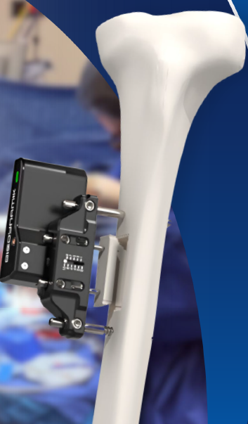
BioDynamik XT3 System XT3 System mounted on tibia

XT3 System

BioDynamik, Inc. — Foothill Ranch, CA

Automated distraction system for both periosteal distraction and transverse tibial transport. Features a miniaturized, programmable motor that advances 0.031 mm/hour (0.75 mm/day). Used in the first fully automated tibial PD case (Feraru, Tan & Armstrong, IWJ 2026). FDA IDE approved for the SMILE-TTT pivotal trial — the first RCT outside China evaluating TTT for diabetic foot ulcers.

FDA 510(k) Cleared FDA IDE Approved ISO 13485 PD + TTT
  • Automated programmable motor — no manual adjustment needed
  • Mini-rail fixator + distractor assembled together
  • Used for both periosteal distraction and transverse tibial transport
  • First automated PD case: toe pressure rose 22→50 mmHg, wound healed
  • SMILE-TTT pivotal trial: 200 patients, multi-center RCT with Mount Sinai West
  • NSF grant recipient (Dec 2024), ADA Innovation Challenge finalist (2025)

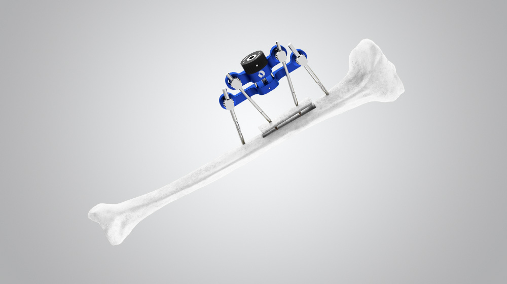
TrueLok Elevate System

TrueLok Elevate

Orthofix Medical Inc.

First commercially available device with a patented design specifically for transverse bone transport (TBT). Two-bar system with half pins and adjustment knob for transverse distraction and compression. Can be used standalone or in hybrid configuration with circular external fixator.

FDA 510(k) Cleared CE Mark TTT
  • Purpose-built for transverse bone transport
  • Half pins D4mm to D6mm
  • Standalone or hybrid with circular fixator (1–2 rings)
  • 130+ procedures completed globally under limited market release
  • Global commercial launch June 2025
  • Active PMCF study: NCT07079774
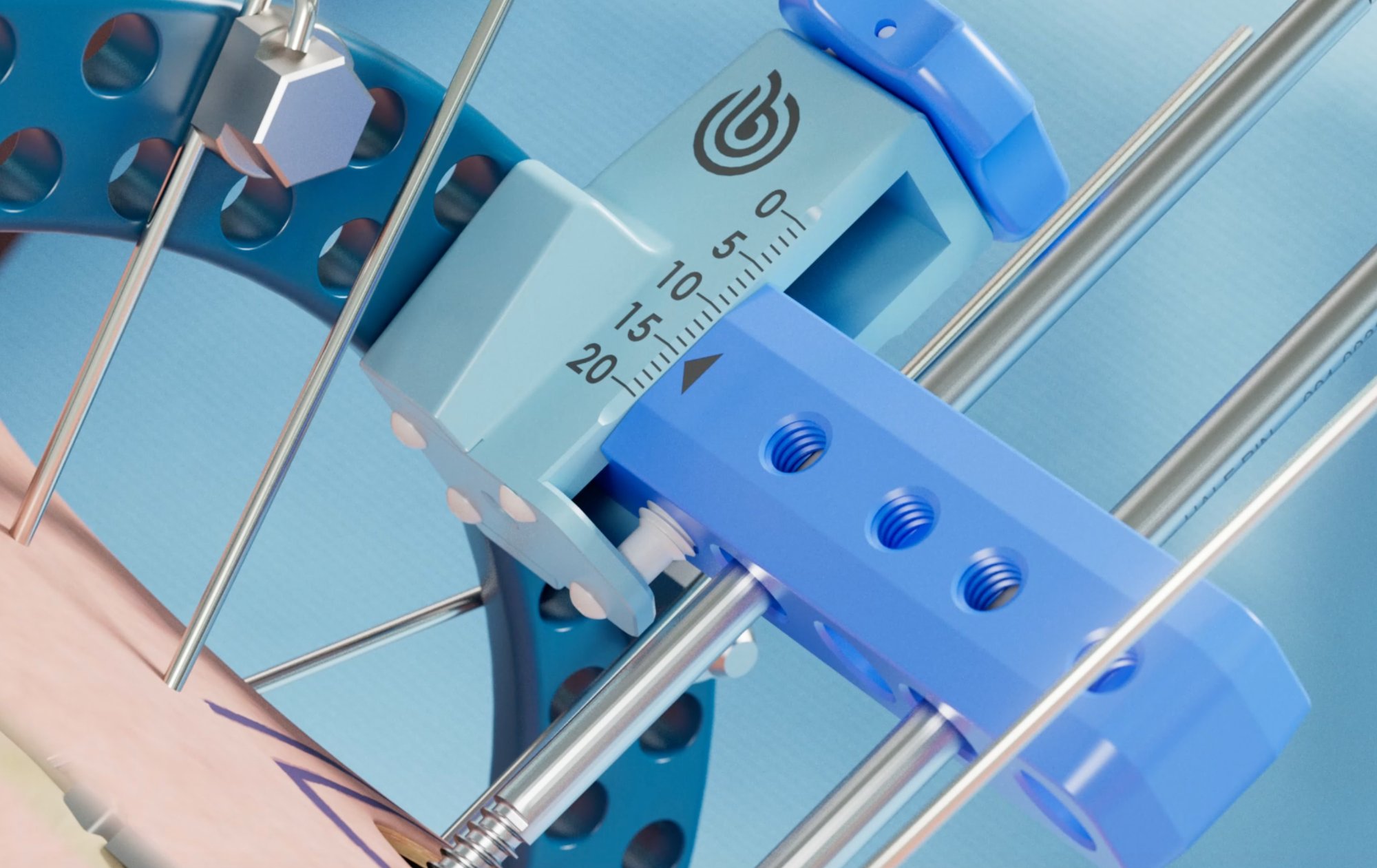
Excelsior External Fixation System

Excelsior External Fixation System

BlueOcean Global Corp.

Next-generation circular external fixation platform designed for limb salvage, incorporating tibial transverse transport (TTT) methodologies advanced from Chinese surgical innovations. Guided by the FASTR (Fixator Assisted Soft Tissue Repair) procedural framework for integrated bone and soft tissue management.

FDA 510(k) Cleared TTT Limb Salvage
  • Modular circular platform for bone transport and reconstruction
  • FASTR framework for staged decision-making
  • Addresses skeletal stability and soft tissue simultaneously
  • FDA 510(k) cleared January 2026
  • Named among "Six Orthopedic Startups to Track in 2026"
  • Surgeon training: FASTR cadaver course (Miami, June 2026)

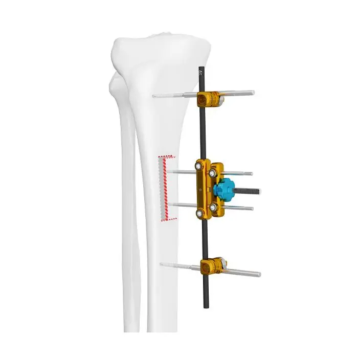
BFIX Transverse Bone Transport System

Aike (Shanghai) Medical Instrument Co., Ltd.

External fixator system specifically designed for transverse tibial bone transport. Compact design with anchor screws for tibial fixation and smaller screws for the cortical bone chip, with a turning nut for inward/outward transport of the bone segment.

FDA 510(k) Cleared CE Mark TTT
  • Purpose-built for TTT — compact unilateral design
  • Two 20 mm anchor screws (1 mm dia.) for tibial fixation
  • Two 10 mm screws (0.8 mm dia.) for cortical bone chip
  • Turning nut for controlled inward/outward transport
  • China Patent 201610722035.5
  • Used in CAOS clinical guideline studies

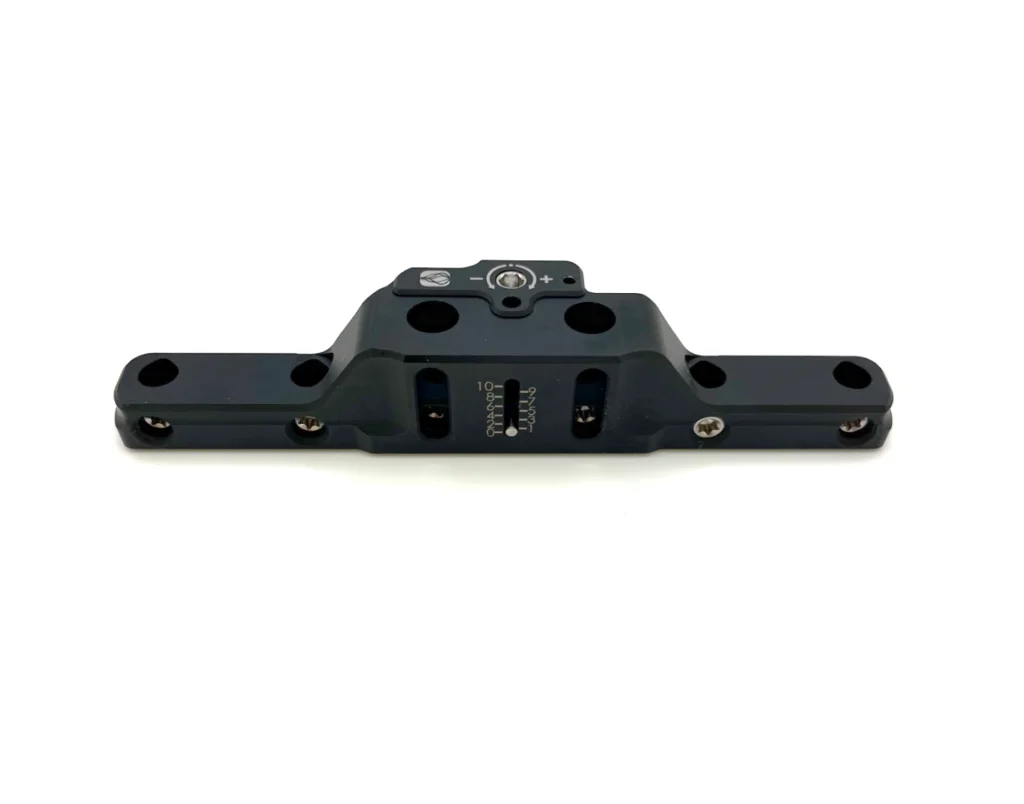
Double Medical horizontal transport external fixator

Modular External Fixator

Double Medical Technology — China

Modular external fixator system comprising threaded pins, adjusting nuts, combination clamps, and carbon fibre rods. Used in clinical research studies and finite element analysis of TTT safety.

CE Mark TTT
  • Modular design with carbon fibre rods
  • Threaded pins + adjusting nuts + combination clamps
  • Used in published TTT research studies

Adaptable Ring & Hybrid Fixation Systems

Multiple manufacturers

Standard Ilizarov-type circular frames and hexapod systems can be configured for TTT off-label. These include well-established platforms from major orthopedic device companies.

FDA Cleared (general fixation) Adaptable for TTT
  • Taylor Spatial Frame (TSF) — Smith+Nephew — hexapod, computer-guided
  • TrueLok EVO & TL-HEX — Orthofix — ring fixation systems
  • Standard Ilizarov frames — multiple manufacturers (JINLU, CZ Meditech, others)
  • Cleared for fracture management; used off-label for TTT
Conferences & Clinical Trials

Where the field is moving — events and active research.

Upcoming Events

June 25, 2026

Mt. Rushmore Diabetic Foot Reconstruction Forum

Sioux Falls, SD — Sanford Research · TTT & PD focus
Details →
August 27–30, 2026

ASAMI-BR & ILLRS 7th World Congress

Bandar Sunway, Malaysia
Details →
September 16–18, 2026

AOFAS & IFFAS Annual Meeting

Foot & ankle orthopedic surgery
Details →
October 22–24, 2026

DFCon 2026

JW Marriott Anaheim, CA — Diabetic foot & limb preservation
Details →
December 5–6, 2026

Diabetic Foot Update (DFUpdate)

UT San Antonio, TX
Details →

Active Clinical Trials

SMILE-TTT: BioDynamik XT3 Pivotal Trial

FDA IDE Approved · 200 patients · Multi-center RCT
BioDynamik.com →

TTT for Chronic Limb-Threatening Ischemia

NCT07175129
ClinicalTrials.gov →

TTT in Management of Chronic Diabetic Wounds

NCT07444593
ClinicalTrials.gov →

TrueLok Elevate PMCF Study

NCT07079774
ClinicalTrials.gov →

Clinical & Mechanistic Study of TTT in Complex Foot Ulcers

NCT05704075
ClinicalTrials.gov →
A Knowledge Network

Independent resources on limb salvage.

Plain-language, evidence-based references for clinicians and patients facing amputation — covering both distraction techniques and the shared goal of saving limbs.

Get In Touch

Questions about the procedure or the evidence?

This is an independent clinical resource. Reach out to discuss the published literature, the mechanism, or how periosteal distraction fits into a limb-salvage pathway.

Periosteal Distraction
© 2026 periostealdistraction.com — Independent clinical resource.
This site provides educational information summarizing publicly available, peer-reviewed literature on periosteal distraction, transverse tibial transport, and related limb-salvage techniques. It is intended for healthcare professionals and informed patients, and is not medical advice or a substitute for clinical judgment. It is not affiliated with, sponsored by, or endorsed by any device manufacturer, and references no specific commercial product. Outcome figures are drawn from the cited studies and may not generalize to all patients or settings. Always consult a qualified clinician about individual care.